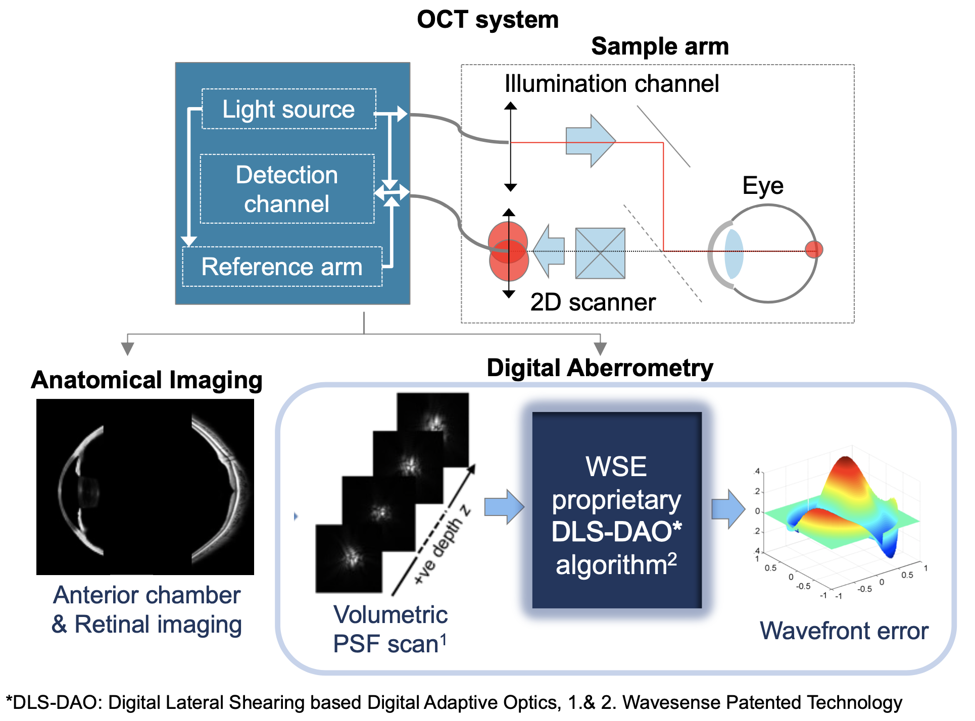
Digital OCT-Aberrometry System is based on Wavesense Patented Technology
Wavesense technology enables OCT system to perform not only anatomical imaging but also Wavefront Aberrometry. The conventional OCT system is adaptable to introduce a separate narrow beam illumination channel, which is derived from the same OCT light source. The illumination beam is directed into the subject’s eye and forms a stationary diffraction-limited spot on the retina, which acts as a pseudo point-source. The light reflected back passes through the full pupil/aperture of the eye and via the same optics and scanners of the sample arm used for conventional OCT imaging and forms the image of the spot at the detection fiber plane. This image of the spot represents the point spread function (PSF) of the eye’s optical system. The scanner laterally translates the PSF at the detection fiber tip resulting in its 2-D sampling. Since it is an OCT system, A-scans are recorded at each translation position. Hence, as a result, a 3-D OCT data recorded, which after OCT based data processing yields a depth resolved 3-D PSF profile of the eye.
The imaged spot corresponding to the photo-receptor layer is selected and processed with our proprietary digital algorithm called Digital Lateral Shearing based Digital Adaptive Optics (DLS-DAO) to calculate the Wavefront Error of the eye.
Multimodal DOCTA system
provides Digital Copy of Patient’s Eye.
Full Eye Imaging provides Anatomical Information.
Aberrometry provides Optical Information.
While state of the art commercial OCT
can only perform Anatomical Imaging,
Wavesense DOCTA system can perform
Wavefront Aberrometry
in addition to
Anatomical Imaging.
Personalized Data and Analysis.
Better Treatment Plan and Care of Patients.
Improved Diagnosis and Surgical outcomes.
Latest Research Articles:
Read more about the latest research based on Wavesense Patented Technology
“Digital ocular swept source optical coherence aberrometry , ” Biomed. Opt. Express (2021)
“Digital adaptive optics based on digital lateral shearing of the computed pupil field for point scanning retinal swept source OCT, “Biomed. Opt. Express 12, 1577-1592 (2021)